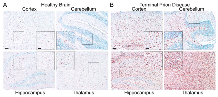
Figure 3.
Microglia are highly activated in terminal prion disease. The pan-microglia/macrophage marker IBA1 is shown in mouse brain sections of (A) mock-infected healthy mice and (B) RML 5.0 prion-infected mice at a terminal disease state. (A) Microglia in the healthy brain show a ramified phenotype with a small soma and thin processes in all four regions displayed here (see magnified close-up). (B) During the terminal prion disease stage, microglia massively proliferate (see overview) and change their morphology towards bigger cell bodies and thicker arms (see close-ups) with a bushy appearance in the hippocampus and an amoeboid phenotype in the thalamus [45,46,47]. Scale bar: 100 µm; close-up: 50 µm.

