Figure 5.
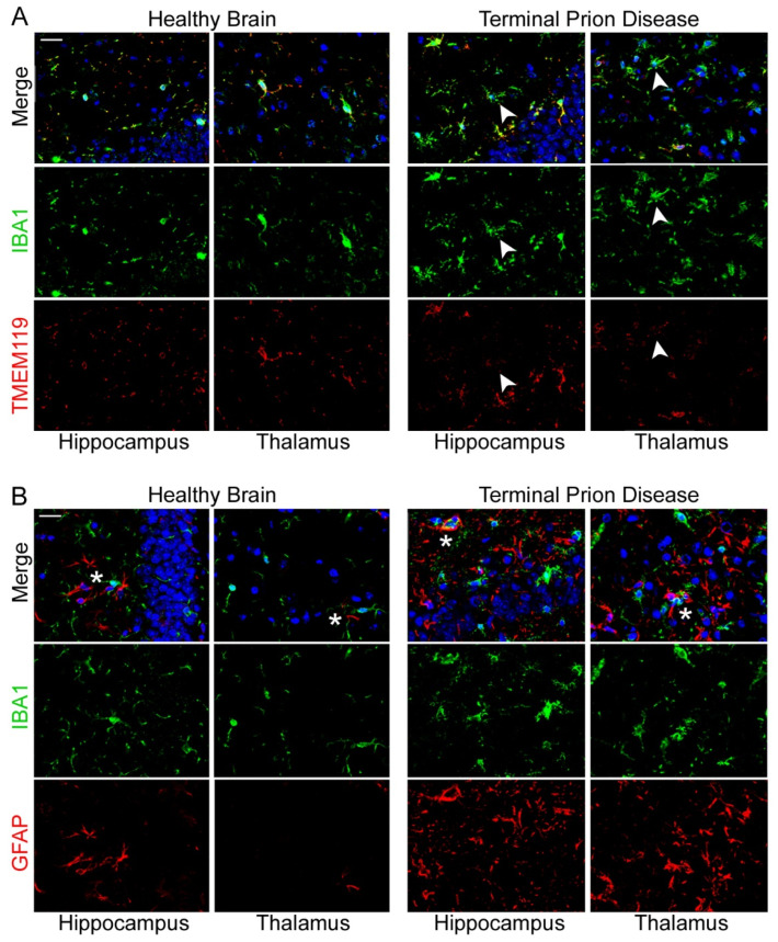
Loss of microglia homeostatic phenotype and increase in gliosis upon terminal prion disease. (A) IBA1 (green) and TMEM119 (red) co-localize in the hippocampus and thalamus in the healthy mouse brain, highlighting the ramified morphology with fine processes mainly stained by TMEM119 [47,73,97]. TMEM119 is highly reduced (white arrowhead) in the dentate gyrus of the hippocampus, and completely lost (white arrowhead) in the thalamus (posterior complex) in terminal prion disease [73]. Scale bar: 20 µm. (B) IBA1 (microglia/green) and GFAP (astrocytes/red) show intense dysregulation and are in close proximity (white asterisks) in the healthy brain and in terminal prion disease. (DAPI/nucleus in blue) [73]. Scale bar: 20 µm.

