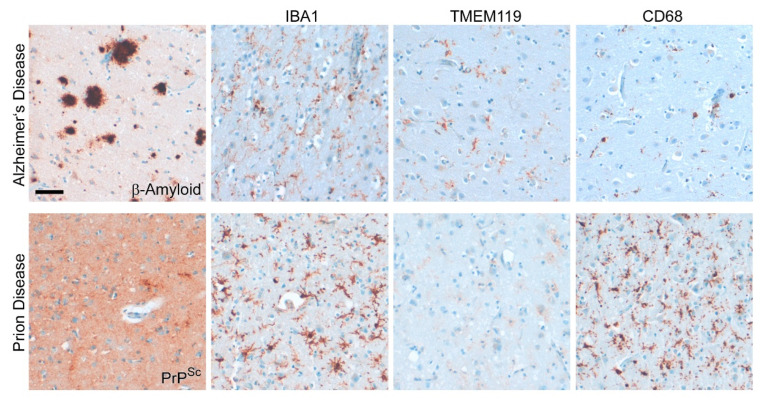
Figure 7.
Disease-associated microglia profile differs between human sCJD and AD. Frontal cortex tissue of a sCJD patient and an age-matched AD patient show reactivity for the pan-microglia/macrophage marker IBA1, the microglial marker TMEM119, and the activation marker CD68 [97]. Deposits of misfolded proteins are shown for the respective antibodies against amyloid-β (AD) or PrPSc (sCJD). Note that the deposition pattern of misfolded PrPSc may vary considerably between CJD patients (see also Figure 6). In contrast to AD, microglia activation is more prominent and the microglial homeostasis marker is almost completely lost in sCJD [97]. Scale bar: 50 µm.

