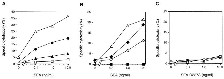
FIG. 2.
Cytokine-treated EC were lysed by SEA-primed T cells, and cytotoxicity was inhibited by specific MAb. (A) SDCC against EA.hy926 cells activated with TNF-α (▴), IFN-γ (•), or a combination of the two cytokines (▵) was compared to that against control cells in the absence of stimulation (□). (B) Cytokine (TNF-α and IFN-γ)-activated EA.hy926 cells were preincubated with MAb (20 μg/ml) directed against CD54 (■) or CD31 (○) and compared to an isotype-matched control MAb (CD10; ⧫). Control cells without any MAb addition were also included (▵). (C) Data are presented from SDCC experiments with the SEA-D227A mutant, which displays a low binding to HLA class II. Symbols are as given for panel A. For panels A and C, EC were treated with IFN-γ and/or TNF-α as indicated in the legend to Fig. 1. EA.hy926 was loaded with 51Cr, and a SEA-reactive T-cell line was used as a source of effector cells at an E:T ration of 40:1. SEA was used at 0.1 to 10 ng/ml. For all panels, the data presented are representative of at least three separate experiments.

