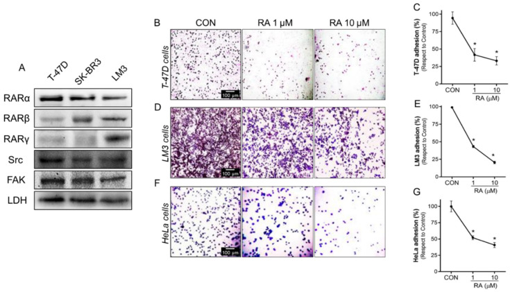
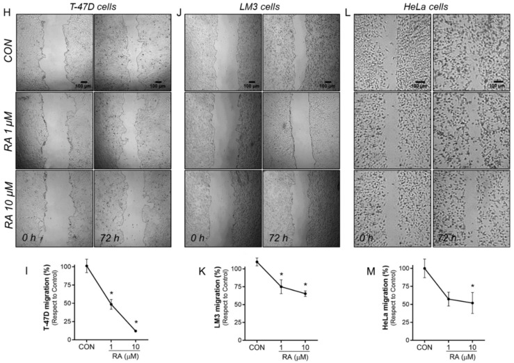
Figure 2.
RA reduces cancer cell adhesion and migration. (A) Whole-cell lysates from human BC cell lines T-47D, SK-BR3, and a murine BC cell line LM3 were evaluated to determine the expression of RARα, RARβ, RARγ, Src and FAK by western blot assay. LDH expression was used as a protein loading control. A representative blot is shown. (B,C) T-47D, (D,E) LM3, and (F,G) HeLa cells were treated with RA (1–10 μM) treatment for 72 h. After the treatment, cells were seeded into 96-well plates previously covered with gelatin and a cell adhesion assay was performed. Representative images and percentages of adhered cells are shown. (H,I) T-47D, (J,K) LM3 and (L,M) HeLa cells were exposed to RA (1–10 μM) for 72 h and cell migration by wound-healing assay was performed. Representative images are shown. Gap closure was quantified by the use of the ImageJ software. Adhesion and migration values are presented as a percentage of control. * = p < 0.05 vs. control, CON. All experiments were performed in triplicates with consistent results.


