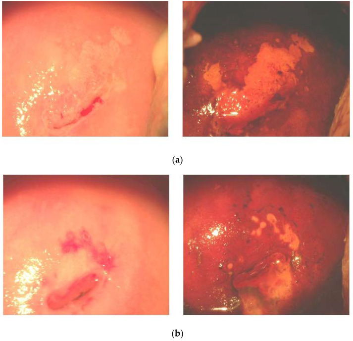
Figure 5.
Typical colposcopy of CIN I and CIN II lesions before (a) and after (b) administration of antioxidants. (a) Before administration of antioxidants (ectopic transformed areas, squamous cell metaplasia, and mosaics marked by the iodine-negative areas). (b) After a three-month course of antioxidants (normal tissue basis, shrinkage of iodine-negative areas, and cervical coagulation). Left panels are colposcopic images without iodine staining. Right panels are colposcopic images after iodine staining.

