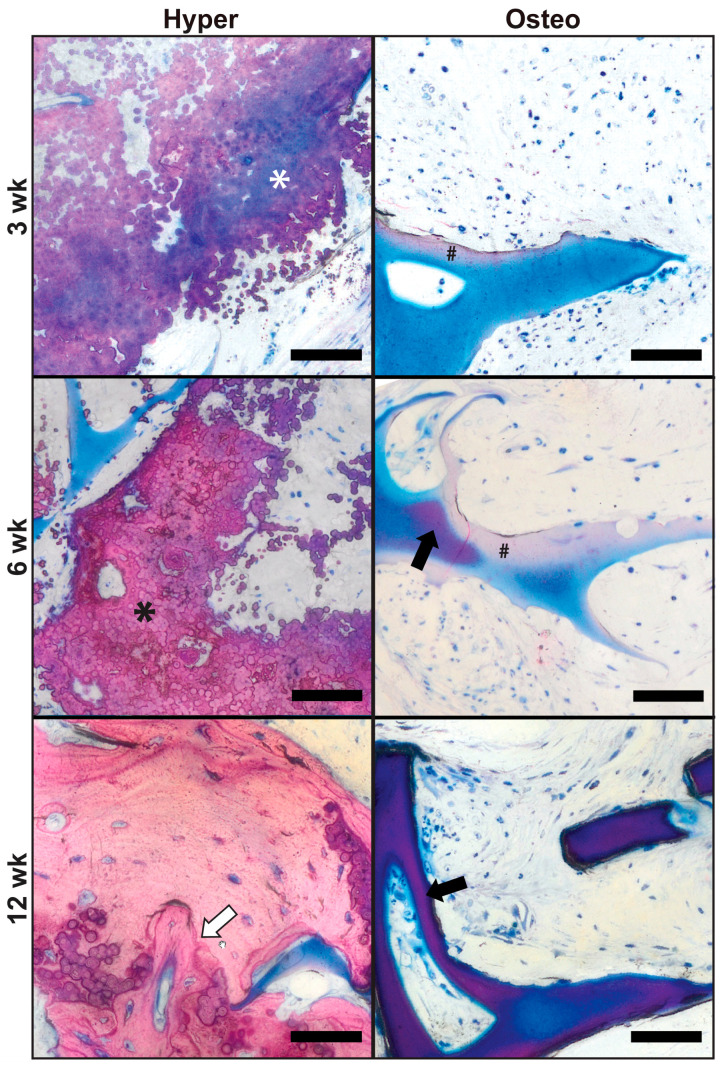
Figure 4.
In vivo bone regeneration. The undecalcified bone histology of harvested constructs was conducted following 3, 6 and 12 weeks of implantation. The samples were stained with Levai–Laczko stain to evaluate the presence of mineral and bone regeneration. In the hyper constructs, calcified nodular deposits exhibiting a blue/purple tint (white asterisk) were present at 3 weeks. Continued mineralization and compaction of the extracellular matrix nodules resulted in a change in the tint to a pink color (black asterisk) in the hyper samples at 6 weeks. Mature bone remodeling was indicated by the presence of a cement line (white arrow) in the hyper samples at 12 weeks. In the osteo samples, mineral deposition was initiated in the margins of the silk scaffold, as seen in the samples at 3 and 6 weeks as light pink staining (black hash sign). Dense scaffold mineralization in the osteo samples was present in limited amounts at 6 weeks (dark purple stain, indicated by the black arrow) and was more widespread at 12 weeks, consistent with the µCT evaluation. Scale bars: 50 µm.

