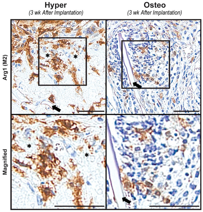
Figure 5.
Presence of macrophages. Arg1 immunohistochemistry staining was conducted to evaluate the presence of M2 macrophages in the constructs after 3 weeks of implantation. Representative images demonstrate numerous M2 macrophages, as indicated by the brown staining localized around the matrix resembling calcified cartilage (the white, round matrix marked by black asterisks) in the hyper constructs. In the osteo constructs, fewer M2 macrophages were found to be localized in proximity to the scaffold (the silk scaffold is indicated by black arrows in both constructs). Scale bars: 50 µm.

