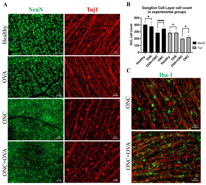
Figure 3.
Immunofluorescence staining of whole mounted retinas. (A) NeuN- and Tuj1-positive cells in the GCL of whole mounted retinas. Scale bar: 50 μm. (B) Statistical analysis of GCL cell count, ns: not significant, *: p < 0.03; ****: p < 0.0001; Welch’s t test. (C) Iba-1 (green) cell infiltration merged with Tuj1 (red). Scale bar: 50 μm.

