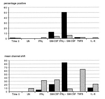
FIG. 1.
Effects of anticoagulants and cytokines on HLA-DR expression on PMN. Heparin and EDTA whole-blood cultures (solid and stippled bars, respectively) were stained immediately (time zero) or incubated for 18 h at 37°C with either PBS (UN), IFN-γ, GM-CSF, IFN-γ plus GM-CSF, TNF-β, or IL-6 and then stained for HLA-DR as described in Materials and Methods. Results are representative of experiments using two donors and are expressed as the percentage of HLA-DR-positive cells and shift in fluorescence intensity (mean channel shift).

