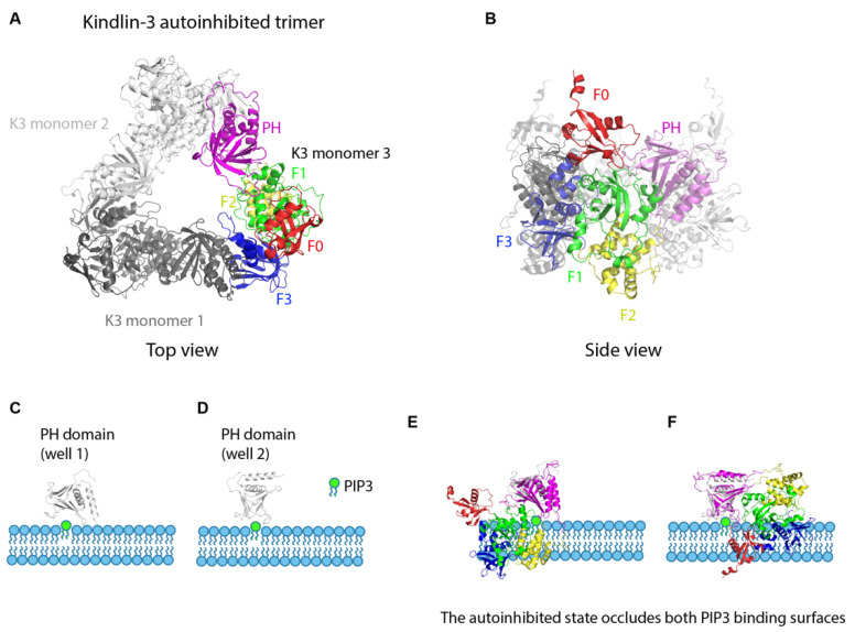
Figure 3.
Kindlin autoinhibition and activation. (A). Crystal structure of the kindlin-3 autoinhibited homotrimer (top view). Kindlin-3 PH domain (magenta) interacts with the F3 subdomain of another kindlin-3 molecule (grey), thereby masking the integrin binding site of kindlin-3. F0, red, F1, green, F2, yellow, F3, dark blue. (B). Crystal structure of the kindlin-3 autoinhibited homotrimer (side view). (C,D). Two wells [well 1 (C) and well 2 (D)] of the free kindlin-3 PH domain binding to PIP3 (green) in the plasma membrane. (E,F). The autoinhibited state occludes both PIP3 binding surfaces of the PH domain. Superimposition of the autoinhibited kindlin-3 monomer onto the kindlin-3 PH domain in (C,D) shows that at least 2 domains would clash with the membrane containing PIP3.

