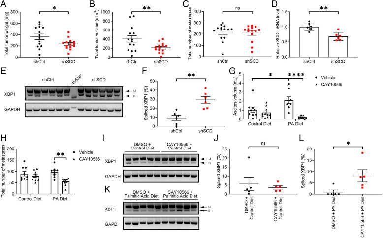
Fig. 6.
SCD KD inhibited growth of OC xenografts in mice. (A) Total tumor weight, (B) total tumor volume, and (C) total number of metastases in athymic nude mice intraperitoneally injected with OVCAR-5 cells transduced with control shRNA (shCtrl) or shRNA targeting SCD (shSCD) and evaluated after 28 d (values are means ± SE, n = 14 per group). (D) qRT-PCR measurements of SCD expression (mean ± SD, n = 6) in a random sample of tumor xenografts described in A. (E) Agarose gel electrophoresis of XBP1 splicing products and (F) percentage spliced XBP1 isoform estimated by image analysis of transcript bands (mean ± SE, n = 6) in a random sample of tumor xenografts described in A. (G) Ascites volume and (H) total number of metastases in athymic nude mice intraperitoneally injected with OVCAR-5 cells, fed with a PA-rich diet or control diet, and treated with SCD inhibitor CAY10566 or vehicle for 28 d. Values are means ± SE, n = 10. (I–L) XBP1 splicing products (I and K) and percentage of intensity of the spliced transcript estimated by image analysis (J and L) in a random sample (n = 5) of tumor xenografts described in F. Values are means ± SE. *P < 0.05, **P < 0.01, ****P < 0.0001.

