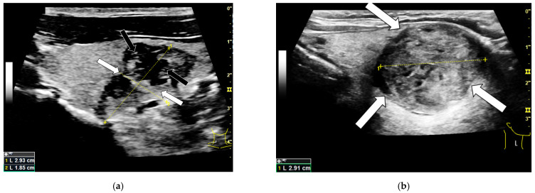
Figure 1.
B-mode examination of a right thyroid lobe with histo-pathologically proven thyroid carcinoma (a) and a histo-pathologically proven thyroid adenoma (b). The carcinoma shows typical malignancy criteria like a blurry edge demarcation (white arrows) as well as an irregular sonomorphological structure and microcalcifications (black arrows). Additionally the majority of the malignoma can be described as hypoechoic compared to the surrounding thyroid tissue. The adenoma shows a clear edge demarcation (white arrows) and a quite homogeneous structure compared to the carcinoma. It is partly isoechoic and partly hyperechoic.

