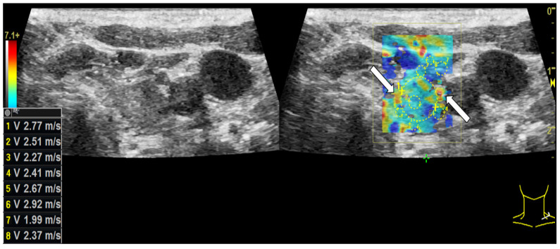
Figure 3.
Shear-wave elastography [m/s] examination on a left thyroid lobe with thyroid adenoma. Only few hard areas (white arrows) can be detected in shear-wave elastography. The major part of the nodule appears rather soft. Neither the stiffness values of the lesion nor of the normal thyroid tissue surpass the calculated cut-off value of 4.04 marginally and 3.88 centrally.

