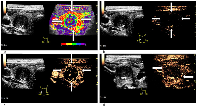
Figure 4.
Patient with thyroid carcinoma. In the parametric colour-coded CEUS mode (a) the margins of the lesion (white arrows) are mainly covered in red and orange. The center of the lesion as well as the normal thyroid gland are primarily coloured in purple with small parts of blue and green. This suggests that SonoVue first expands along the carcinoma’s margins. The CEUS examination 5 s after the bolus injection (b) shows no contrast agent expansion yet neither marginally (white arrows) nor centrally. 9 s after the bolus injection (c) the contrast enhancement has begun: Microbubbles first expand along the lesion’s margins (white arrows). In the CEUS recording, taken 2 min after bolus injection (d), a decrease of contrast enhancement called wash-out along the carcinoma’s margins (white arrows) can be spotted.

