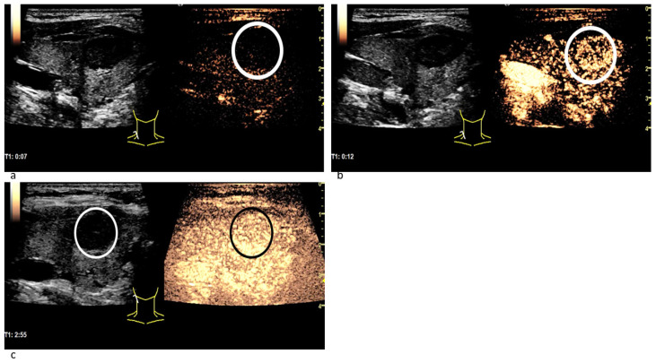
Figure 5.
Patient with a benign thyroid nodule. The CEUS examination 7 s after the bolus injection (a) shows no contrast agent expansion in the nodule (white circle), yet. 12 s after the bolus injection (b) the contrast enhancement has begun: Microbubbles expand inside the lesion quite homogeneously (white circle) and not from the margins to the center. In the CEUS recording taken almost 3 min after bolus injection (c) the contrast agent intensity has not decreased: neither in the nodule (white circle in B-mode; black circle in CEUS) nor in the surrounding thyroid tissue. No wash-out phenomenon has occurred.

