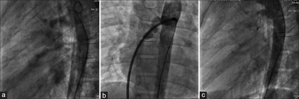
Figure 1.
(a) Descending aortogram in left lateral view showing the fistulous tract opacifying the right atrium and right ventricle. (b) Sheath injection in the fistula in anteroposterior projection after formation of arterio-venous loop. (c) Descending aortogram after deployment of duct occluder within the fistula

