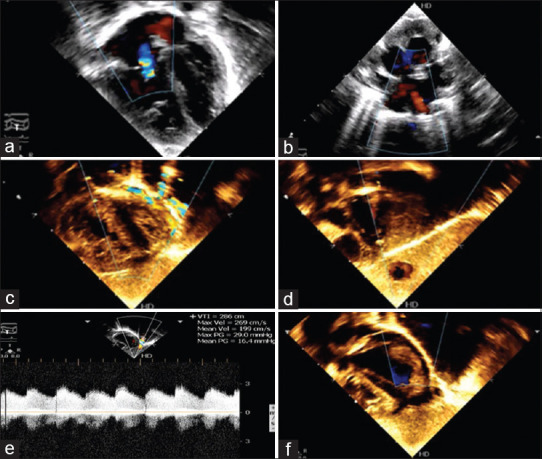
Figure 2.

(a) Echocardiogram (4-chamber view) showing dilated right heart and mild tricuspid regurgitation. (b) Modified short axis view showing at least two pulmonary veins draining normally to the left atrium. (c) Subcostal 4-chamber view showing the fistulous tract with colour, running lateral to the left ventricle. (d) Subcostal view showing dilated portal vein with colour flows. (e) Doppler interrogation across the fistula showing restriction (gradient: 29/16 mm Hg) as it enters the diaphragm. (f) Echocardiogram (4-chamber view) post device closure of the fistula does not show any colour flow in the area lateral to the left ventricle
