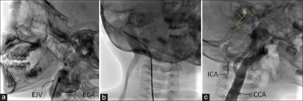
Figure 4.
(a) Contrast injection by hand through Judkins right diagnostic catheter in the right external carotid artery shows the tortuous fistulous communication between the external carotid artery and the right external jugular vein. Image obtained after turning the head to the left lateral position. (b) Contrast injection through the Judkins right guide catheter after deploying ADO-II in the terminal portion of external carotid artery just before its communication with the external jugular vein, shows filling of the dilated right external jugular vein and faint filling of the medial right internal jugular vein. (c) Contrast injection in the right common carotid artery after device release (device marked by the yellow box bracket) shows dilated common carotid artery and unobstructed flow to the right internal carotid artery. Image obtained after turning the head to the right lateral position

