Figure 7.
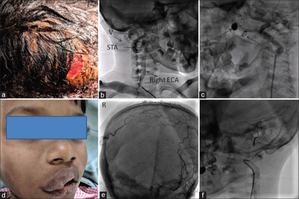
(a) Scalp arteriovenous fistula showing scalp erosion. (b) Contrast injection through Judkins right diagnostic catheter in the right external carotid artery showing a dilated external carotid artery and superficial temporal artery and immediate opacification of venous tributaries (v) of the external jugular vein. (c) Left external carotid artery shoot showing a mildly dilated left external carotid artery and coils in situ, suggestive of contra-lateral filling of the scalp arteries. Image obtained after turning the head to the right. (d) The huge scalp arteriovenous fistula has caused swelling of the right face and lips. (e) Left external carotid artery injection (Towne view) showing contralateral filling of the superficial scalp plexus. Right side has been marked “R.” (f) Contrast injection via end-hole catheter placed deep within the left external carotid artery showing opacification of the left occipital artery and coil in the left posterior auricular artery

