Figure 1.

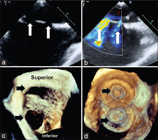
Two-dimensional transesophageal echocardiogram in vertical plane (a) shows superoinferior orientation of two defects (white arrows) with color flows (b). Three dimensional right atrial enface view in anatomical orientation (c) shows the superoinferior orientation of the two defects (black arrows) before and after (d) device closure
