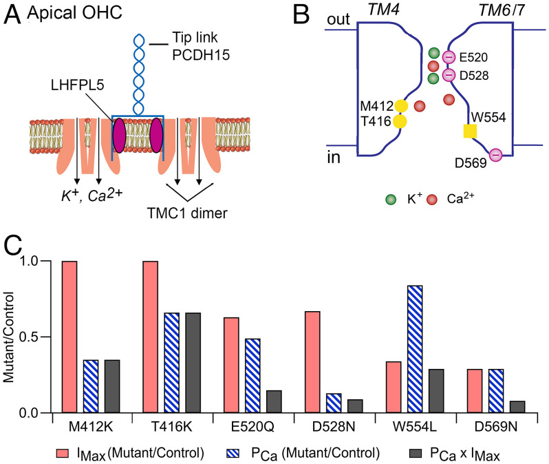
Fig. 6.
Schematics of OHC transducer complex and TMC1 channel. (A) At the cochlear apex, two dimeric TMC1 channels are shown in the stereociliary membrane. Each dimer is attached to an LHFPL5 molecule, which in turn is connected to one PCDH15 of the tip-link. Pore locations indicated by arrows, each transporting K+ and Ca2+. Not shown are TMIE subunits thought to be linked to TMC1 channel or the CIB2, which is assumed to attach on the inner face of the TMC1. (B) Hypothetical cross-section of the channel formed by TM domains 4, 5, 6, and 7, exhibiting an outer vestibule, a neck region (E520, D528), and a larger inner vestibule (M412, T416). W554 on inner face of TM6 and D569 on cytoplasmic link to TM7 may interact with LHFPL5. Mutations discussed in the text nullify the negative charges (E520Q, D528N, D569N) or add a positive charge (M412K, T416K), to increase the electrostatic barrier for transmission of positively charged cations. (C) Effects of six homozygous Tmc1 mutations on the peak MET current (IMX), Ca2+ permeability of the MET channel (PCa), and Ca2+ influx into the hair bundle (IMX × PCa). Both IMX and PCa are expressed as mutant/control.

