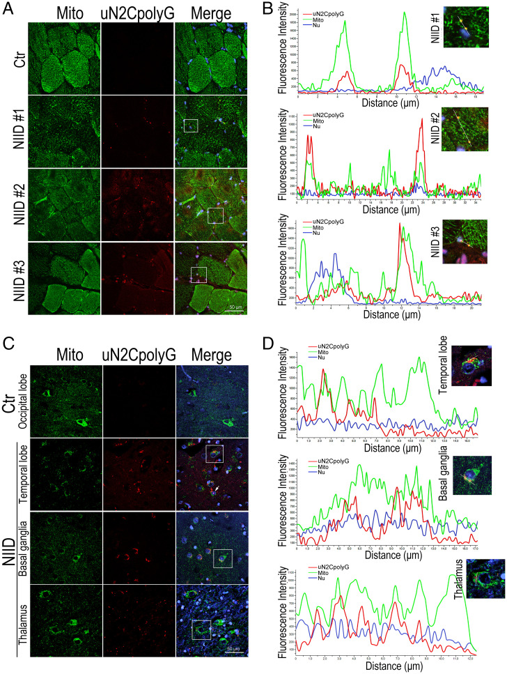
Fig. 4.
Colocalization of uN2CpolyG and mitochondria in skeletal muscle and brain from NIID patients. (A) Representative immunofluorescence microscopy images of skeletal muscle samples from three NIID patients and one normal control, which reveals the distribution of uN2CpolyG in muscle fibers. uN2CpolyG protein partially colocalized with mitochondria. (B) Line scan analysis of the merged images shows uN2CpolyG was colocalized with mitochondria in muscle fibers from NIID patients. (C) Representative immunofluorescence microscopy images of brain samples from one NIID patient and one normal control, which reveals the distribution of uN2CpolyG in brain samples. uN2CpolyG protein partially colocalized with mitochondrial. The arrow indicates uN2CpolyG-positive intranuclear inclusion. (D) Line scan analysis of the merged images shows uN2CpolyG was colocalized with mitochondria in three different brain regions (temporal lobe, basal ganglia, and thalamus) from the NIID patient. Scale bar, 50 μm (A and C).Nu, nucleus.

