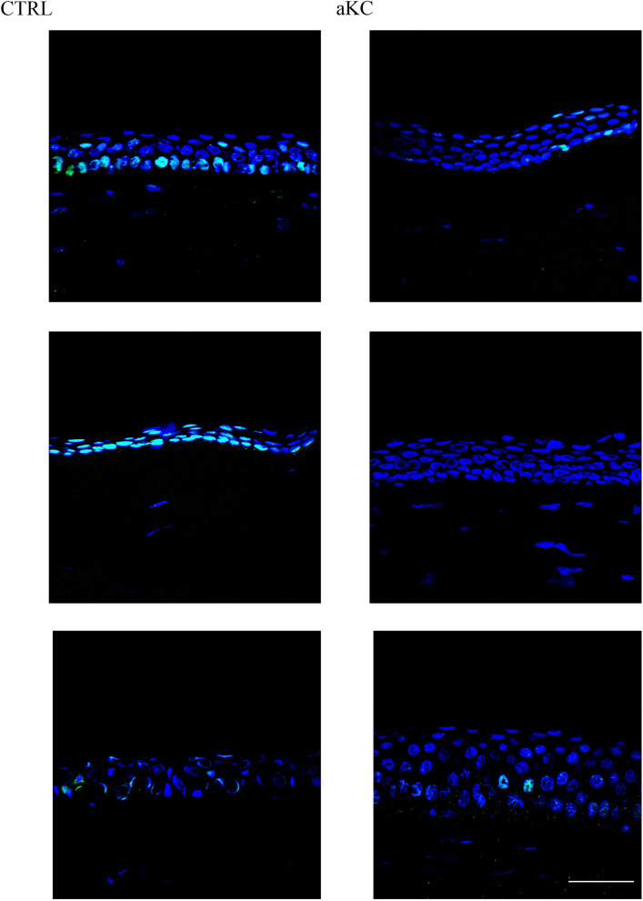
Fig 4. NRF2 active form staining in human corneal material.
Sections of normal (CTRL; n = 5) and advanced keratoconus (KC; n = 3) corneas were analyzed by indirect immunofluorescence with an antibody targeting the active phosphorylated form of NRF2 (green). Nuclei were stained with TO-PRO Iodide (blue). Scale bar = 100 μm. Figure show three representative controls and keratoconus.

