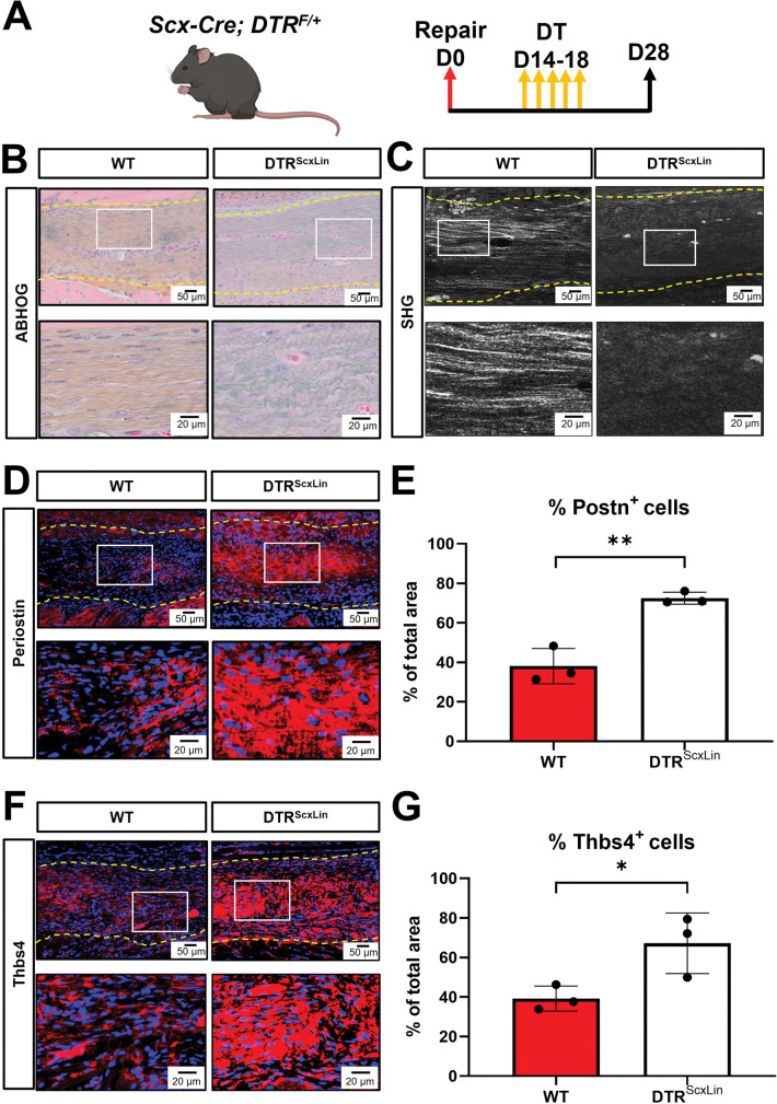
Fig 5. DTRScxLin tendons exhibit an immature bridging matrix tissue at D28 post-surgery.
A. Schematic of the mouse model used and timeline for tendon surgeries, DT injections, and tissue harvesting. B. ABHOG staining to visualize the structure and organization of the healing DTRScxLin vs WT tendons. C. SHG imaging to visualize mature collagen fibrils in the healing DTRScxLin vs WT tendons. D. Periostin staining (red) of D28 DTRScxLin vs WT tendons. E. Quantification of Periostin+ immunostaining area in the D28 DTRScxLin vs WT tendons. F. Thrombospondin 4 staining (red) of D28 DTRScxLin vs WT tendons. G. Quantification of Thrombospondin 4 + immunostaining area in the D28 DTRScxLin vs WT tendons. Nuclei are stained with DAPI (blue). N = 3 per genotype.

