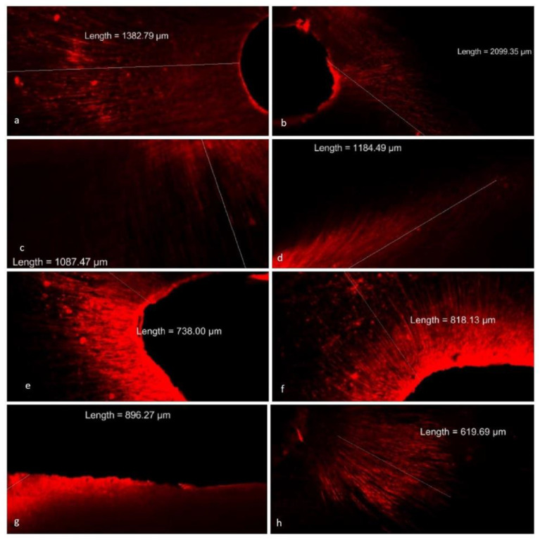
Figure 4.
Confocal images depicting the depth of penetration of liposomal CHX containing rhodamine B dye into dentinal tubules. Photographs (a,b) depict the penetration of liposomal CHX into dentinal tubules in coronal third. Photographs (c,d) show liposomal CHX penetrating in middle third. (e,f) Liposomal CHX average penetration in apical third was less when compared with other thirds. (g,h) Images of penetration in the coronal and apical third, respectively of normal CHX. The penetration was significantly less as compared with liposomal CHX.

