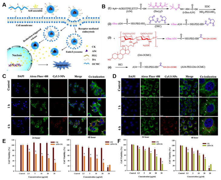
Figure 6.
(A) Structural pathway of APD-CK inside the cell. (B) Conjugation of polymer (A54-PEG-DA-OCMC) before loading ginsenoside CK. Cellular uptake of APD-CK nanoparticles after incubating for 1 h and 6 h with (C) HepG2 and (D) Huh-7 cells. (E) In vitro cytotoxicity of normal ginsenoside CK and APD-CK against HepG2 cell line. (F) In vitro cytotoxicity of normal ginsenoside CK and APD-CK against Huh-7 cell line. Asterisks (*) denotes statistical significance difference compared with control group. Reprinted with permission from Ref. [117]. 2020, Elsevier Ltd.

