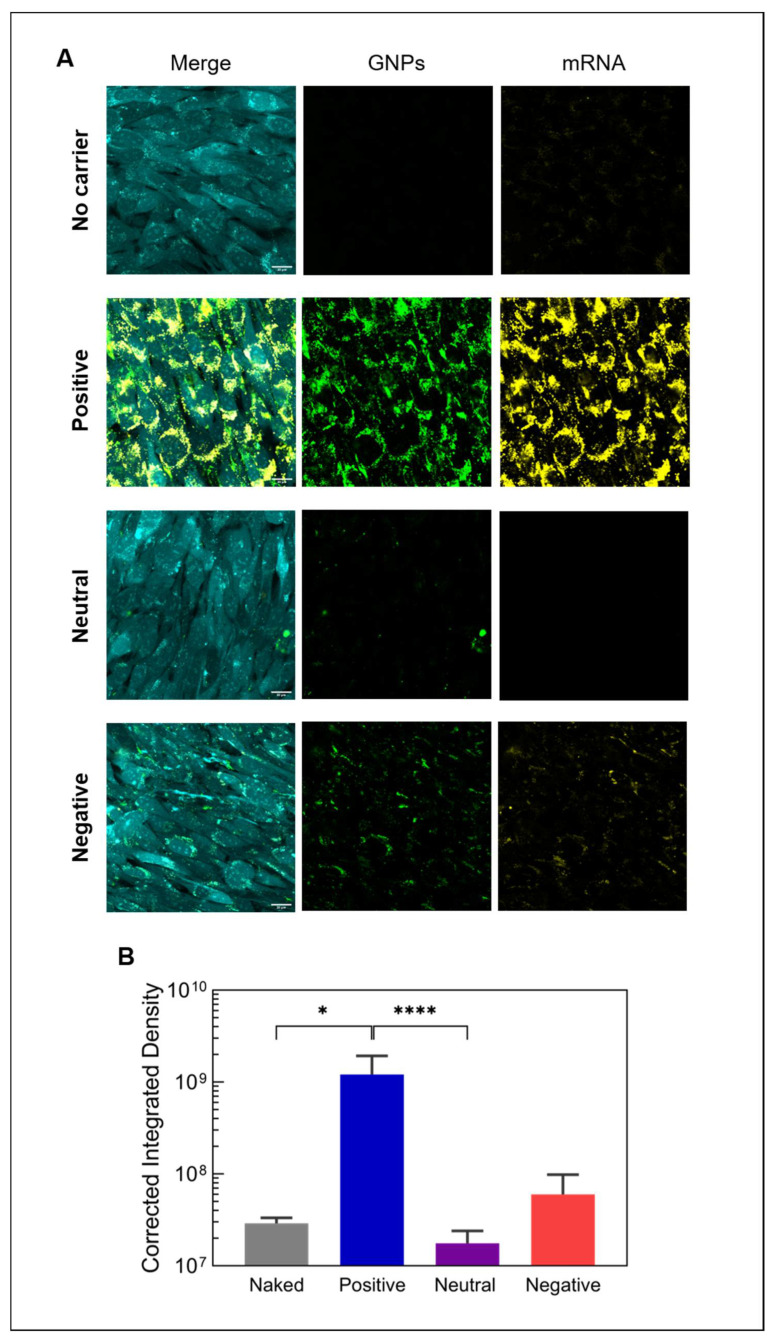
Figure 5.
Internalization of mRNA-loaded gelatin nanoparticles. (A) Confocal live cell images showing the uptake of fluorescently labeled mRNA (yellow) by mouse pre-osteoblastic cells (cyan) after 24 h using differently charged gelatin nanoparticles (green) and (B) the quantification of internalized mRNA (n = 6). Naked mRNA refers to mRNA delivered without any vector or particle (no carrier). All images were acquired using the same acquisition settings. Scale bar represents 20 µm. Statistical significance is shown as * p < 0.05 and **** p < 0.0001.

