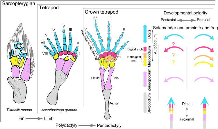
Fig. 1. Structural homology between sarcopterygian fins and tetrapod limbs, with comparison of previous understandings on limb developmental sequences during early and late skeletogenesis in salamanders versus that in frogs and amniotes.
Note that distal carpals/tarsals 1 and 2 remain separate in the schematic hindlimb of crown tetrapods, but only a single element (basale commune) is present in salamanders and some early tetrapods. Following Gegenbaur’s scheme (2, 73), the mesopodium in the schematic hindlimb of crown tetrapods is divided into three transverse rows in different colors to highlight homologous structures that are present or absent in fish fins: distal (red), central (orange), and proximal (yellow). The main text generally follows Goette’s scheme (see definition in Materials and Methods) (74) of dividing the mesopodium into three columns for the ease of developmental analyses. Previous understandings of the developmental patterns of tetrapod limb structures along the anteroposterior and proximodistal axes are denoted by corresponding, color-coded arrows (right figure). Acanthostega hindlimb (7) and Tiktaalik pectoral fin (9). c, centrale; dt, distal tarsal; f, fibulare; i, intermedium; t, tibiale; y, element y.

