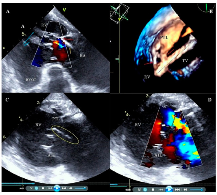
Figure 4.
Adhesion of the lead to the tricuspid apparatus. Broken chordae tendineae during transvenous lead extraction (TLE). (A) 2D transesophageal echocardiography (TEE) transgastric view, color Doppler. The ventricular lead (yellow arrow) is fused with the posterior leaflet (red arrow) and the subvalvular apparatus. Papillary muscles (blue arrows). Low tricuspid regurgitation. (B) 3D TEE. Tricuspid valve (TV) view from the right ventricle (RV) side. Tissue bridges (adhesions) (red arrows) connecting the ventricular lead (dashed line) with the posterior leaflet (PTL). (C) 2D TEE transgastric view. Broken chordae tendineae (circle) that moves to right atrium (RA) (D) 2D TEE transgastric view. Image from panel C in the option of color Doppler, large TR to RA, Vena contracta (VC) = 11 mm (black arrow).

