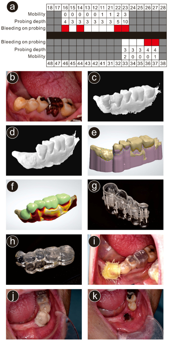
Figure 2.
Case presentation of the participant in this report. (a) Periodontal chart at the first visit. Gray-colored blocks indicate missing teeth and red-colored blocks indicate bleeding on probing; (b) intraoral view before extraction; (c) STL data obtained from the intraoral scanner (TRIOS 3); (d) stereolithography data after deletion of the left mandibular canine; (e) designing the surgical splint using three-dimensional (3D) computer-aided design software; (f) designed surgical splint; (g) the surgical splint printed using a 3D printer; (h) trimmed and finished surgical splint; (i) delivered surgical splint; (j) intraoral view 2 days after the extraction (before removal of the surgical splint); (k) intraoral view of the extraction wound 2 days after the extraction.

