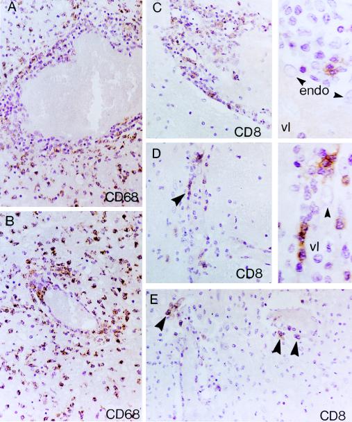
FIG. 3.
Patient no. 1 (A and B) Distribution of CD68+ monocytes/macrophages in TVE involving medium-sized (A) and small-sized (B) cerebral vessels. (A) A zonal angiocentric pattern of inflammation characterized by a rim of CD68− TVE and, to a lesser degree, PV lymphocytic infiltrates, surrounded by an outer zone consisting predominantly of CD68+ monocytes/macrophages. (B) A small vessel exhibiting focal TVE involvement by CD68+ monocytes/macrophages. The intervening brain neuropil (A and B) is dominated by heavy accumulations of CD68+ cells. (C to E) Distribution of CD8+ lymphocytes in TVE involving medium-sized (C) and small-sized (D and E) vessels. (C) Distribution of CD8+ cells in TVE and PV. Note the distinct predilection of CD8+ lymphocytes for the inner portions of TVE in juxtaposition to the endothelial lining (arrowheads) (see also right insets in panels C and D). Also note the paucity of CD8+ T cells in the intervening neuropil. endo, endothelial cells; vl, vessel lumen. ABC immunoperoxidase and counterstaining with H&E were used for all panels. Original magnifications, ×400 (A to D) and ×1,000 (insets in panels C and D).

