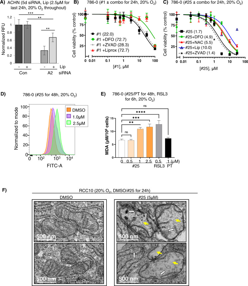
Fig. 5. ISCA2 inhibition induces ferroptosis.
A Effect of co-treatment of ISCA2 siRNA and DMSO or liproxstatin (2.5 μM) on cell viability determined using resazurin in ACHN cells. siRNA transfection was performed for a total of 5 days with liproxstatin (or DMSO) added for the last 24 h. B, C Cell viability assays (resazurin) of 786-0 cells treated with (B) #1 or (C) #25, ± DFO (100 μM), liproxstatin (1 μM) or ZVAD-FMK (20 μM); or (H) #25 ± DFO (100 μM), NAC (1 mM), liproxstatin (1 μM) or ZVAD-FMK (20 μM). Treatments were performed for 24 h. Average IC50 values (µM) are shown in brackets. D Impact of #25 treatment (48 h) on BODIPY 581/591 fluorescence detected by flow cytometry in 786-0 cells. E Impact of 48 hours’ treatment with #25, PT2385 (PT) or 6 hours’ treatment with RSL3 on malondialdehyde (MDA), an indicator of lipid peroxidation in RCC10 cells. F Transmission electron microscopy of RCC10 cells treated with DMSO or #25 for 24 h (2 representative micrographs of each condition). DMSO-treated cells show normal mitochondria whereas #25-treated cells show damaged mitochondria including lost or irregular cristae (white arrows) and irregular matrix with voids (yellow arrows).

