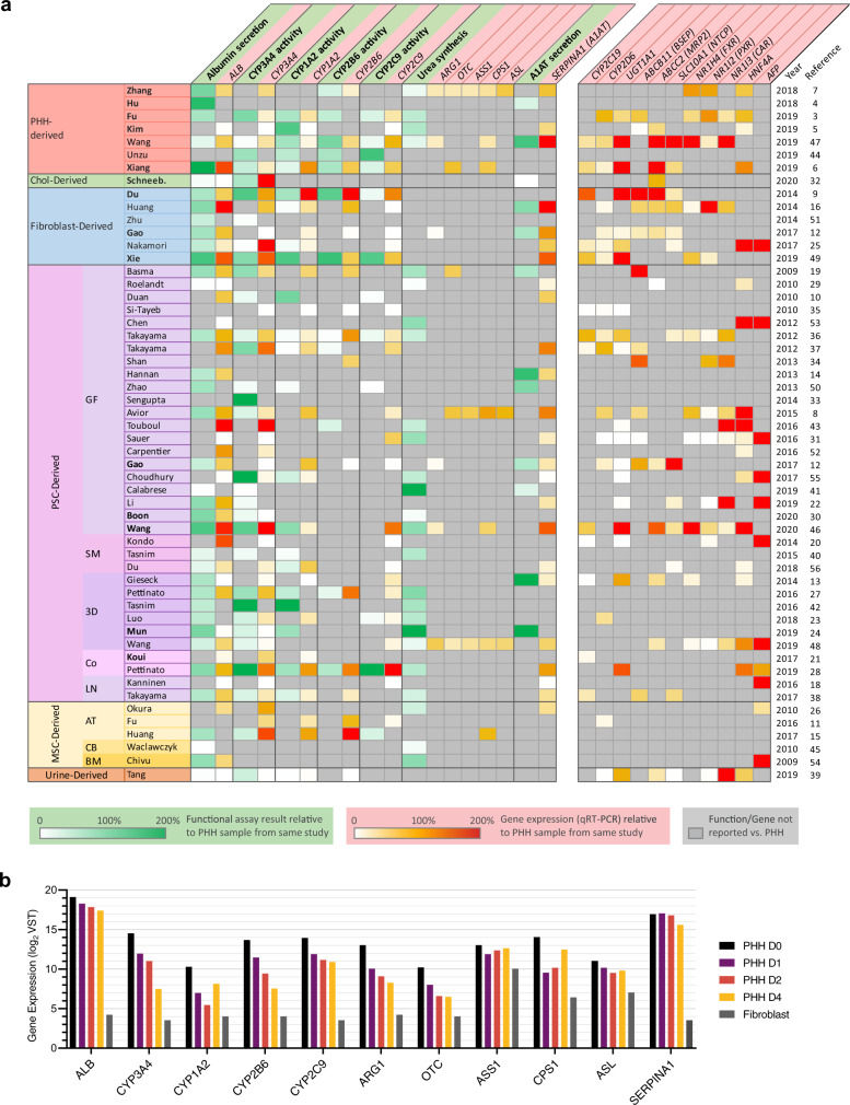
Fig. 1. Reported hepatic functional assays and gene expression are insufficient for HLC comparison.
a The most commonly reported hepatic functions and gene expression are presented. The value in each cell represents the activity or expression level of an HLC as a percentage of the PHH control in the same study. HLCs are grouped based on the type of cells they were generated from. Pluripotent stem cell (PSC) derived HLCs are further grouped into: GF, standard protocols employing growth factors; SM, protocols solely using synthetic molecules; 3D, protocols utilizing 3D matrices; Co-cult, protocols combining multiple cell types; and LN, protocols focusing on the effect of laminin coating. Mesenchymal stem cell (MSC) derived HLCs are categorized into the tissue of origin: AT adipose tissue, CB cord blood, BM bone marrow. b mRNA expression (log2) of genes relevant to the commonly reported hepatic functions in PHHs cultured for 0, 1, 2, and 4 days and cultured dermal fibroblasts, from the study of Gao et al.12. Note that expression of some genes (e.g. CYP3A4, CYP1A2, OTC) decreases >4-fold within the first 24 h of culturing, whereas expression of other genes (ALB, ASS1, SERPINA1) remains relatively stable. Source data are provided in Supplementary Data 3.

