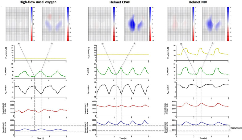
Fig. 1.
Comparison of representative tracings of airway pressure, transpulmonary pressure esophageal pressure and global and regional electrical impedance tomography during spontaneous breathing with high-flow nasal, helmet CPAP and NIV in a patient with severe hypoxemic respiratory failure. The left panel shows the respiratory mechanics during spontaneous breathing with high flow oxygen mask. Due to the high inspiratory effort and to the inhomogeneity of the lung, it is possible to appreciate the Pendelluft effect. The start of inspiration (marked by the initial negative deflection of the Pes) is coincident with the increase of electrical impedance tomography in the Global ROI tracing (∆Z, %). However, while in the dorsal regions of the lungs (dependent regions) there is an increase of ∆Z%, in the ventral region there is a decrease of ∆Z% (non-dependent regions). This represents the “Pendelluft effect”, an intra-tidal displacement of air from non-dependent to dependent lung regions, causing local overstretch of the latter. The first dotted line marks the moment when the ∆Z% signal in the most ventral ROI stops decreasing and local inflation begins. In right panels, the respiratory mechanics of the same patient receiving helmet CPAP and pressure support are shown. High PEEP generates recruitment in dorsal lung regions and mitigate the pendelluft effect and enhances more homogeneous lung inflation. Presence of pressure support causes a decrease of the inspiratory effort ∆Pes swing. Heat maps describe lung regional inflation (blue pixels) and deflation (red pixels). In the absence of PEEP, a significant pendelluft effect is documented (red pixels during inspiration), which reflects the intra-tidal shift of gas from anterior non-dependent lung regions to posterior dependent lung regions. This is abolished by high PEEP delivered through the helmet interface, which makes inflation homogenous across the whole lung tissue. Acronyms: PAW, airway pressure; PES, esophageal pressure; ∆Z %, electrical impedance tomography signal variation; ROI, region of interest; VV, ventral-ventral; MV, middle-ventral; MD, middle-dorsal; DD, dorsal-dorsal

