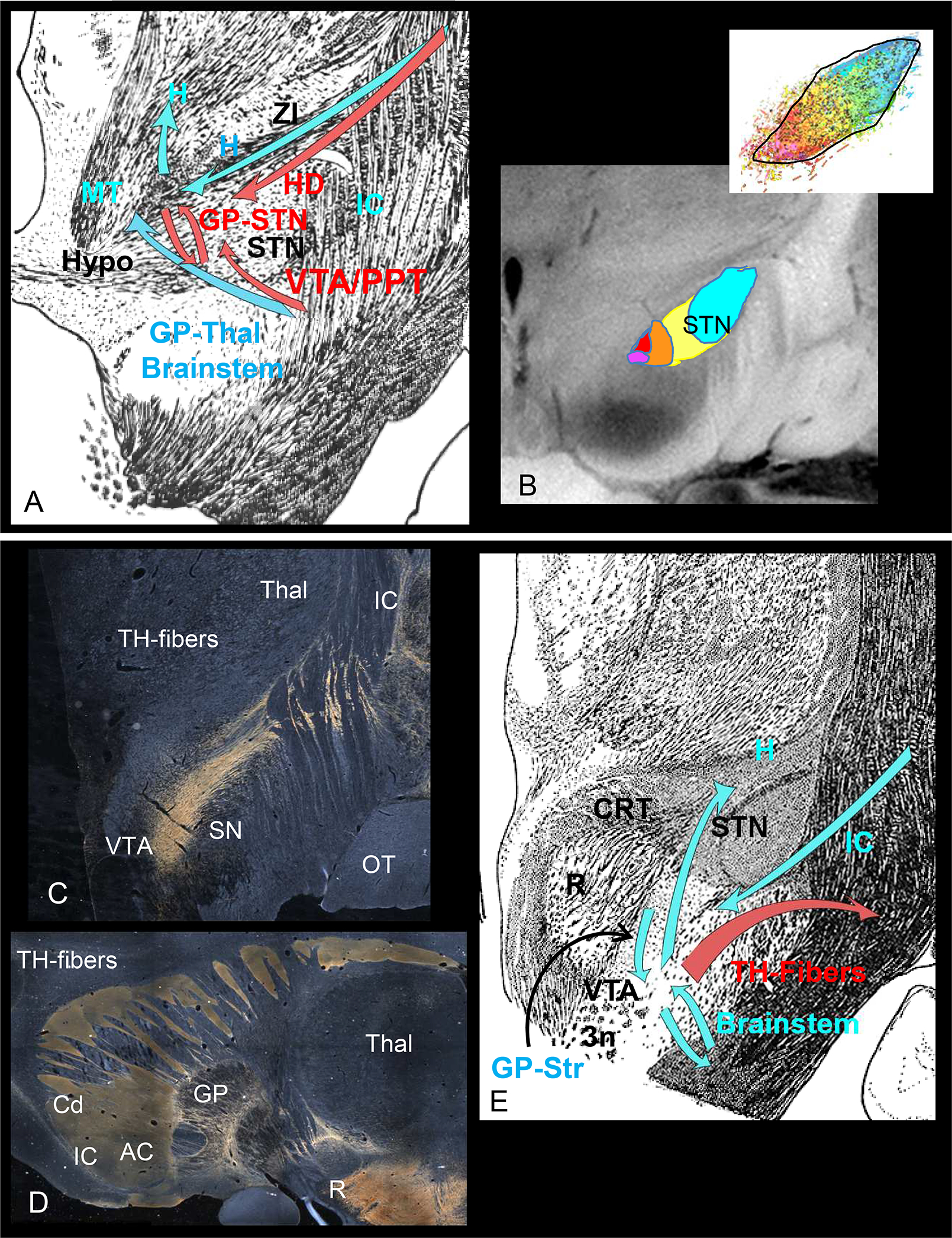
Figure 2.

Connections through the STN(A/B) and midbrain (C/D) sites. A. Fiber bundles and connections through the STN. Red arrows=targeted connections, blue arrows=other pathways through the area. B. Organization of cortical terminals in the STN. Color code: Fuchsia=vACC/mOFC, red=OFC, orange=dACC, yellow=dPFC/vlPFC, blue= premotor/motor cortex. C. Dark field illumination demonstrating TH-positive fiber-positive staining (appears gold) as they travel from the VTA laterally, cross the IC to terminate in the globus pallidus and striatum. D. Sagittal section demonstrating the trajectory of TH-positive fibers to frontal cortex. Note, the TH-positive fibers are only found in the striatum, but not in the IC. E. Fibers bundles and connections through the midbrain site. Red arrows=targeted connections, blue arrows=other pathways through the area. Abbreviations: AC=anterior commissure, ALIC=anterior limb of the internal capsule, Cd=caudate, GP=globus pallidus, H=H fields of Forel, HD=hyperdirect pathway, Hypo=hypothalamus, IC= internal capsule, MT=mammillothalamic tract. PPT=pedunculopontine nucleus, OT=optic tract, R=red nucleus, SN=substantia nigra, STN=subthalamic nucleus, TH=tyrosine hydroxylase, Thal=thalamus, VTA=ventral tegmental area, ZI=ZI, 3n=third nerve. Scale bar=5mm.
