4.

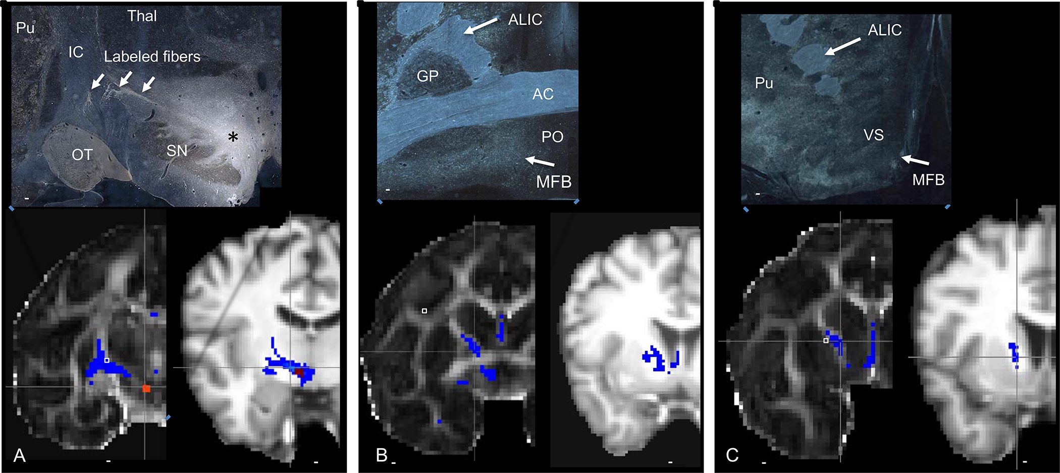
Projections from the VTA. Top panels= NHP histology following a tracer injection into the VTA, bottom panels, left=NHP dMRI tractography in NHP, right= human dMRI tractography. Top panel. A. asterisk=tracer injection site, Red=seed placement at the same site as the injection in monkey and human dMRI. Similar to the anatomic tracing, streamlines cross the IC, to the striatum. However, unlike the anatomic tracing experiment, streamlines also enter the IC and continue to travel rostrally, through the ALIC. B-C. Histology demonstrates fibers in the MFB, ventral and medial to the anterior commissure. There are no fibers in the ALIC. In contrast, the dMRI tractography in both the monkey and human show streamlines in the ALIC. Abbreviations: AC=anterior commissure, ALIC= anterior limb of the internal capsule, GP=globus pallidus, IC=internal capsule, MFB=medial forebrain bundle, OT=optic tract, PO=preoptic area, SN=substantia nigra, VS=ventral striatum. Scale bars: top panel-5mm; bottom panel, NHP-4.9mm; human-10mm.
