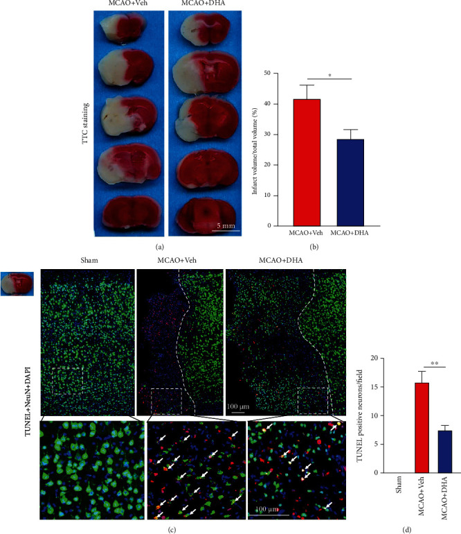
Figure 2.

DHA reduced the infarct volume and alleviated neuronal death after ischaemic stroke. (a) Representative images of TTC-stained brain sections in each group (scale bar =5 mm). (b) Quantification of the infarct volume in each group. (c) Representative fluorescence images of cells co-labelled with TUNEL and NeuN in the ischaemic penumbra in each group (scale bar =100 μm). White dotted lines separate the normal and penumbra regions, and white dotted squares demarcate the enlarged area. White arrows indicate neurons co-labelled with TUNEL and NeuN. (d) Number of TUNEL-positive neurons per field in the infarct area. Data are expressed as the mean ± SEM (n =6 animals from each group) (∗, P < 0.05; ∗∗, P < 0.01). The two-tailed Student's t-test (b) and one-way ANOVA followed by Tukey's post hoc test (d) were used for data comparison.
