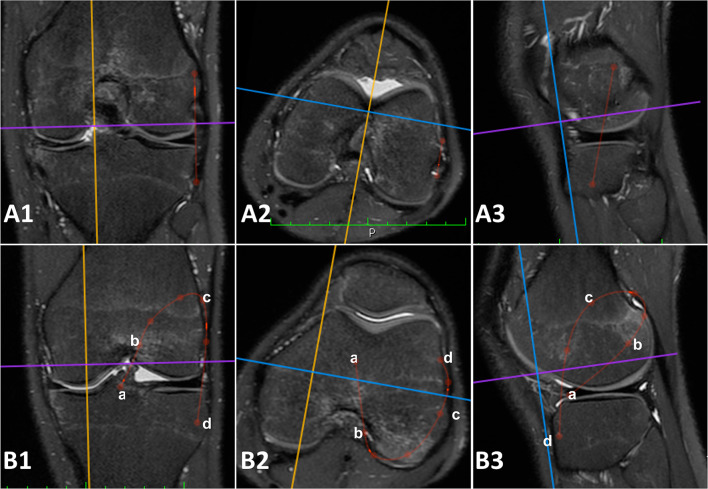
Fig. 3.
3D measurements performed on the 3D MRI sequences (Turbo Spin Echo T2 weighted 3D MRI with isotropic voxel). A Red lines: anatomic anterolateral ligament (ALL) reconstruction defined as the distance from a point located 5 mm proximal and 5 mm posterior to the apex of the lateral femoral epicondyle and a tibial point equidistant between the center of the Gerdy’s tubercle and the anterior margin of the fibular head, 9.5 mm distal to the joint line. Particularly, A1 is the coronal view, A2 is the axial view, and A3 is the sagittal view of a left knee with simulation of the same reconstruction. B Red lines: Marcacci technique (MT) defined as defined as the ACL graft path of the Marcacci technique, passing through a the center of the tibial ACL footprint; b the over the top femoral position; c the distal aspect of the proximal ridge of the femur at the diaphyseal-metaphyseal junction; d the Gerdy’s tubercle. Particularly, B1 is the coronal view, B2 is the axial view, and B3 is the sagittal view of a left knee with simulation of the same reconstruction

