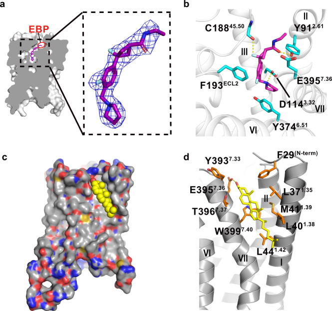
Fig. 2. Binding modes of PF-03654746 and cholesterol to H3R.

a Vertical cross section showing a shallow binding pocket in H3R. The extending binding pocket (EBP) of H3R-PF03654746 is shown in a red ellipse. |2Fo|−|Fc| electron density map for the PF-03654746 contoured at 1.0σ. b Detailed interactions of PF-03654746 in the H3R ligand-binding pocket. H3R was shown in gray ribbons, with critical residues for ligand-binding as cyan sticks and PF-03654746 as magenta sticks. Hydrogen bonds were shown as yellow dashed lines. c Surface representation of cholesterol-binding site with cholesterol shown in yellow spheres. d Detailed interactions of cholesterol with H3R. Residues critical for cholesterol binding were shown as orange sticks and cholesterol was shown as yellow sticks. The hydrogen bond was shown as yellow dashed lines.
