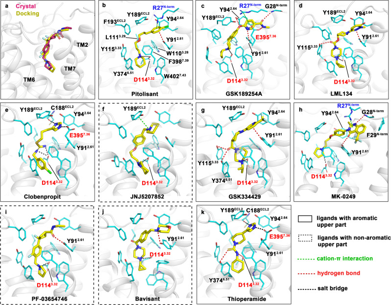
Fig. 3. Docking results of PF-03654746 and nine different H3R antagonists.
a Superimposition of PF-03654746 in the crystal structure (magenta) and the docked pose (yellow). b–k Binding modes of 10 H3R antagonists. The docked ligands were depicted as yellow sticks and arranged according to their Ki values from low to high as shown in Supplementary Table 2. Ligands with an aromatic moiety in the upper part of their binding poses were boxed with solid lines and ligands with non-aromatic upper parts were marked with dash lines. Interacting residues were presented as cyan sticks. In b, all residues involved in interactions were labeled. In c–k, only residues forming interactions with the external aromatic moiety or involved in polar contacts were labeled. Charged residues were marked in red (negative) or blue (positive). Polar interactions, including cation–π interactions, hydrogen bonds, and salt bridges, were represented by different colored dash lines, while π–π stackings were not shown.

