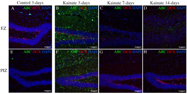
Figure 5.
Immunohistochemical evidence for canonical Wnt activation in the hippocampus. Immunofluorescent images demonstrating active beta-catenin (ABC—a marker of canonical Wnt pathway activation) and doublecortin (DCX—a marker of immature dentate granule cells) in (A–D) the EZ (upper row), and (E–H) the PIZ (lower row) dentate gyrus. Images demonstrate active beta-catenin expression level (green) (A,E) in control mice 3 days after saline injection (7 days and 14 days unchanged, data not shown), and in kainate-injected mice (B,F) 3 days, (C,G) 7 days, and (D,H) 14 days after SE. Scale bar 100 µm.

