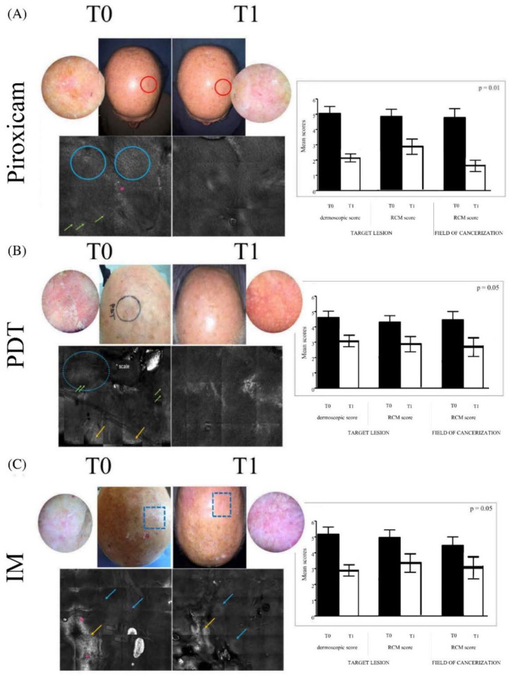
Figure 1.
Clinical, dermoscopic, and reflectance confocal microscopy evaluation before (T0) and after treatment (T1). In the panels on the left, the images represent the clinical aspects of the target lesion and the field of cancerization of the scalps of different patients treated with different therapies: (A) medical device 0.8% piroxicam and 50+ sunscreen, (B) photodynamic therapy (PDT), (C) 0.015% ingenol mebutate (IM) gel; the dermoscopic features of the target lesion, in detail; and reflectance confocal microscopy mosaic (6 × 6) aspects of the target lesion and the field of cancerization. The blue circles and arrows indicate the atypical honeycombing pattern, constituted by pleomorphic keratinocytes; the red asterisks areas of detached keratinocytes; the green arrows inflammatory infiltrate; and the yellow arrows hyper- and para-keratosis. In the panels on the right, we report the dermoscopic and reflectance confocal microscopy scores of the target lesion and the reflectance confocal microscopy scores of the field of cancerization for the respective treatments.

