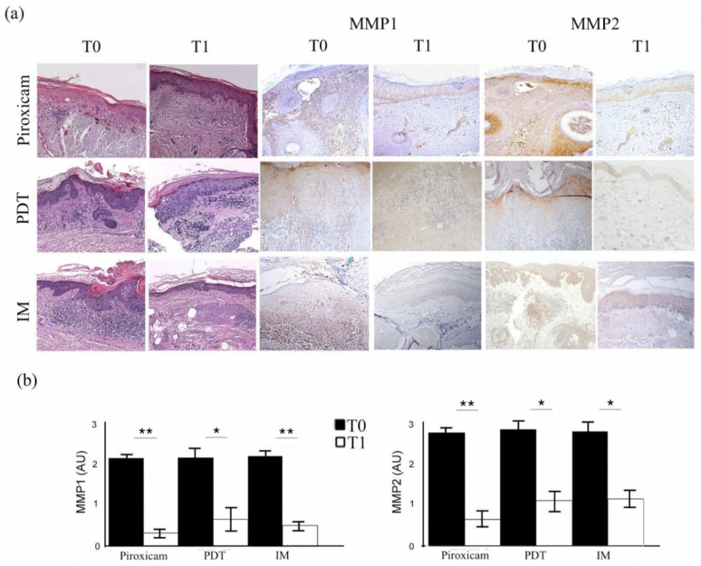
Figure 2.
Histopathological features and metalloproteinase-1 and -2 expression in target lesions before (T0) and after treatment (T1). (a) The first two columns show the histopathological aspects of actinic keratoses before and after treatment with medical device 0.8% piroxicam and 50+ sunscreen, photodynamic therapy (PDT), and ingenol mebutate (IM) gel (hematoxylin–eosin, original magnification: 100×). The two central columns show images demonstrating the immunohistochemical expression of matrix metalloproteinase-1 (MMP1), while the last two show the staining for matrix metalloproteinase-2 (MMP2) in the target lesions before and after each treatment (original magnification: 100×). (b) Semiquantitative evaluation of matrix metalloproteinase expression before and after each therapeutic agent. * p < 0.01; ** p < 0.001.

