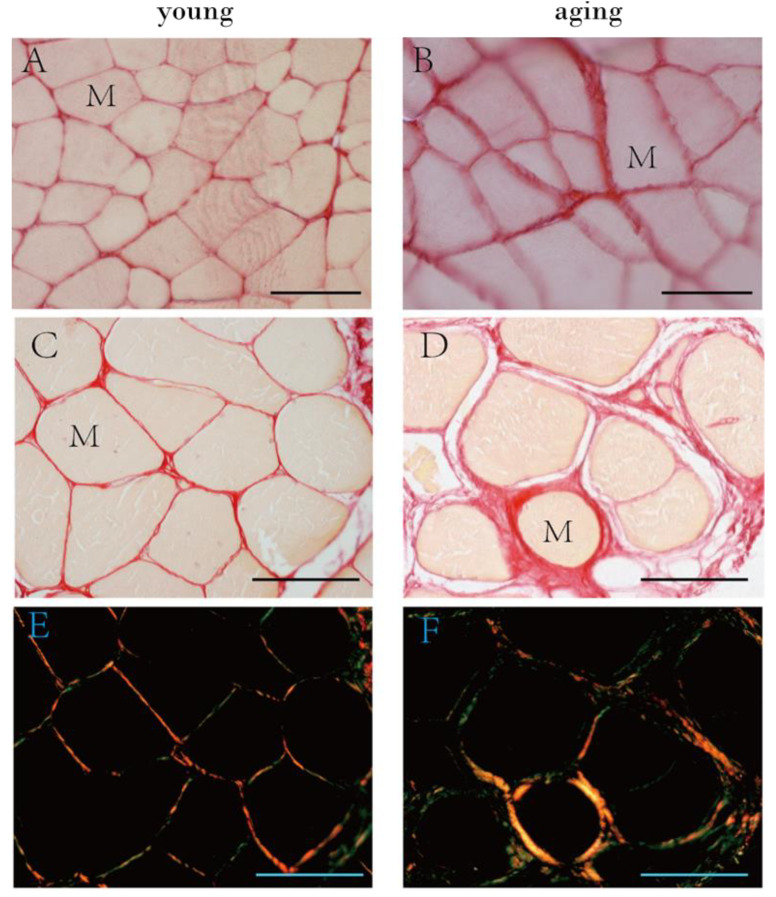
Figure 6.
Picrosirius red staining of mouse (A,B) and human muscle samples (C–F) and polarized light images in human samples (E,F). (A): muscle sample from 6 week old mice (puberty, Group A), (B): from 2 year old mice (elderly, Group C); (C,E): muscle sample from a 32 year old human patient; (D,F): muscle sample from an 87 year old human patient. M = muscle cell. Scale bar: 50 μm.

