Figure 1.
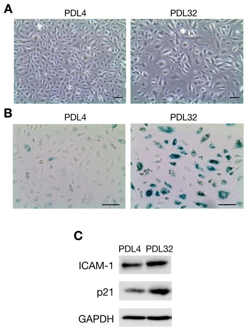
The characteristics of senescent endothelial cells prepared by the serial passage of human umbilical vein endothelial cells (HUVECs). HUVECs were cultured in endothelial cell growth media in 10 cm-diameter dishes and then passaged every 3 or 4 days. (A) Phase contrast images of population doubling level 4 (PDL4) (left) and PDL32 (right) cells are shown. PDL is defined as the total number of times that the cells in the population have doubled. PDL32 cells have an enlarged and flattened morphology (original magnification ×50). (B) Images showing SA-β-Gal staining with PDL4 (left) and PDL32 (right) cells. SA-β-Gal activity is increased in the PDL32 cells (original magnification ×100). (C) The expression of ICAM-1 and p21/WAF-1 in PDL4 and PDL32 cells was evaluated by Western blotting. Images are representative of three independent experiments. Glyceraldehyde 3-phosphate dehydrogenase (GAPDH) expression was detected as an internal control. Scale bars, 100 µm [93].

