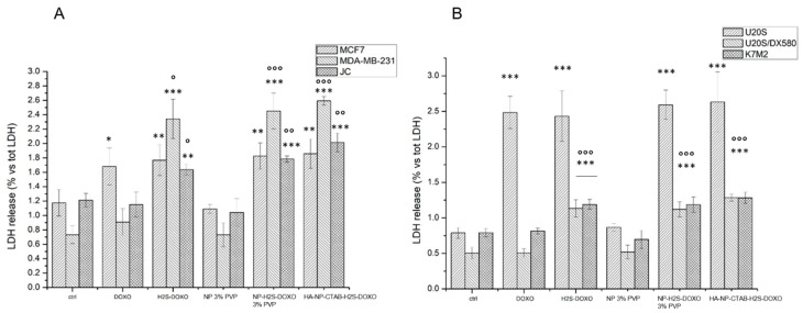
Figure 9.
Acute cytotoxicity in: (A). breast cancer cells; (B). osteosarcoma cells. Cells were incubated 24 h with 5 µM with free DOXO, free H2S-DOXO, NP-H2S-DOXO 3% PVP, and HA-NP-CTAB-H2S-DOXO (containing 5 µM H2S-DOXO), then the release of LDH was measured spectrophotometrically (n = 3). Data are means + SD. * p < 0.05, ** p < 0.01, *** p < 0.001: versus ctrl; ° p < 0.05, °° p < 0.01, °°° p < 0.001: versus DOXO.

