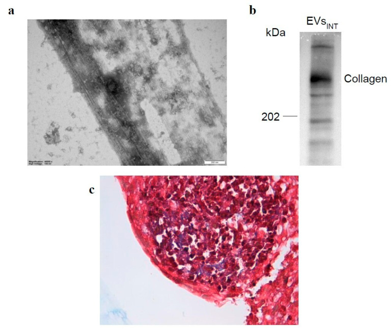
Figure 5.
Collagen in EVsINT sample. (a) Ultrastructural TEM image of EVINT sample highlighting the presence of fibers; the size bar is 200 nm. (b) Western blotting of collagen in EVsINT sample. (c) Masson trichrome stain (conventional staining procedure) most likely showing, in blue/purple, the presence of connective tissue; image 60×.

