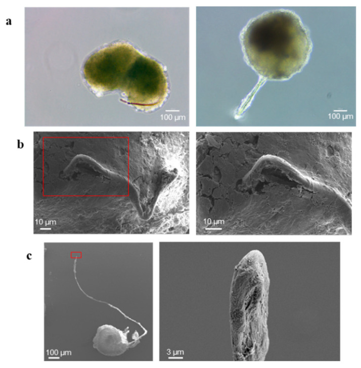
Figure 6.
Vasculogenic mimicry-like phenomenon. (a) Images of some representative spheroids containing tubules observed by optical microscopy; the size bar is 100 µm. (b,c) SEM images of representative spheroids presenting tubules: (b) shows the tubule exiting from the spheroid; (c) shows the tip of the tubule; each red rectangle encloses the microscope field shown, in higher magnification, on the image on the right, highlighting the cavity of the tubule. The size bar is 10 µm in (b), 100 and 3 µm in (c), in order from the left to the right image.

