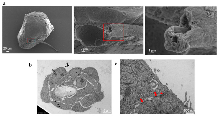
Figure 7.
Lumen inside the vasculogenic mimicry-like tubules. (a): SEM images highlighting the tubule lumen; each red rectangle encloses the microscope field shown, in higher magnification, on the image on the right, highlighting the cavity of the tubule. The size bar is 20, 2 and 1 µm, in order from the left to the right image. (b): TEM image of a sagittal section of the tubule, highlighting the hollow lumen delimited by cells. The size bar is 5 µm. (c): representative TEM image showing the cellular junctions: the arrows point to tight junctions, the arrowhead points to desmosome. The size bar is 500 nm.

